
Conditions & Treatments General Urology Understanding Kidney Stones Treatment Options for Large Kidney ... Percutaneous Nephrolithotomy (PCNL) A Step-by-step Look At The Procedur ...
PCNL Surgery: Step-by-Step
-
 When it is time for your PCNL surgery, a member of the surgical team will
bring you back to the procedure room where you will receive anesthesia.
When it is time for your PCNL surgery, a member of the surgical team will
bring you back to the procedure room where you will receive anesthesia.
- You will be turned, face down on the operating room table (prone position). You will be placed on a specialized bed designed for prone surgery.
- A small tube will be placed from below up to the kidney to allow for precise visualization of the kidney using real-time X-rays.
- Once the location of the stone is mapped, your surgeon will make a small, 1 cm (about 1/2 an inch) incision in the back.
- A small tube is placed into the drainage system of the kidney close to the stone being treated. X-ray imaging called fluoroscopy is used to help guide the precise placement of the tube into the kidney.
- A wire is then passed into the kidney and a balloon is used to dilate a tract.
- A tube is placed over the balloon and a scope is inserted into the tract to look into the kidney.
- Through this tube, the surgeon inserts a small telescope for visualization of the stone as well as specialized instrumentation that is used to break up the kidney stone(s) into very small pieces, which are then cleared from the kidney. The goal is to clear all stones and fragments from the kidney.
- Once the stones are removed, it is not uncommon to place a small tube down the ureter and into the bladder to allow the kidney to drain better while it heals from the procedure.
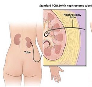
- Rarely, a tube is left in the kidney (called a nephrostomy tube) to allow for additional drainage.
- If necessary, the nephrostomy tube is placed into the surgical site and exits through the skin. This tube is utilized for drainage of urine and blood and is typically removed in about three days following surgery.
- Stones will be sent for analysis to aid in developing a program to prevent further stone formation.