
Types of Vasectomy Reversal
Although a vasectomy is intended as a permanent form of birth control for men, some men’s life circumstances change and they wish to restore their fertility to conceive a child naturally. Vasectomies can be reversed through two primary methods: vasovasostomy and epididymovasostomy.
Vasovasostomy
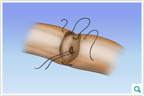 A vasovasostomy is one type of vasectomy reversal procedure that reconnects
the severed ends of the vas deferens. Though there are various techniques
for performing a vasovasostomy, our surgeons prefer a two-layer procedure
that produces the most optimal watertight result when reconnecting the
vas deferens. This is done with microscopic sutures, using the most advanced
microsurgical equipment and the Zeiss operating microscope. The two-layer
procedure involves suturing the mucosal inner layer and a muscular tissue
layer of the vas deferens.
A vasovasostomy is one type of vasectomy reversal procedure that reconnects
the severed ends of the vas deferens. Though there are various techniques
for performing a vasovasostomy, our surgeons prefer a two-layer procedure
that produces the most optimal watertight result when reconnecting the
vas deferens. This is done with microscopic sutures, using the most advanced
microsurgical equipment and the Zeiss operating microscope. The two-layer
procedure involves suturing the mucosal inner layer and a muscular tissue
layer of the vas deferens.
Facilitating the two-layer sutures properly requires a higher level of training and experience, which our surgeons have. It is very important for the closure to be watertight so that sperm do not leak out at the closure site, potentially causing inflammation.
Epididymovasostomy
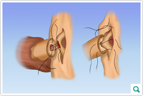 Epididymovasostomy is an even more complex and delicate vasectomy reversal
procedure that is performed when a blockage is discovered in the epididymis.
The epididymovasostomy requires greater surgical expertise that the vasovasostomy.
Epididymovasostomy is an even more complex and delicate vasectomy reversal
procedure that is performed when a blockage is discovered in the epididymis.
The epididymovasostomy requires greater surgical expertise that the vasovasostomy.
The vas deferens is attached directly to the epididymis. An incision is made to an epididymal tubule, where sperm are stored, just prior to the obstruction. The tubule is gently squeezed for fluid. The fluid is checked for sperm. If sperm are absent, a more proximal incision is made and checked again. For epididymovasostomy to be successful, sperm must be present within the tubule at the site of the anastomosis, which is where the sutures will join the two streams (the tubule and the end of the vas deferens). The anastomosis is performed using two layers of very fine sutures and is also done using the operating microscope.